Stop and Go

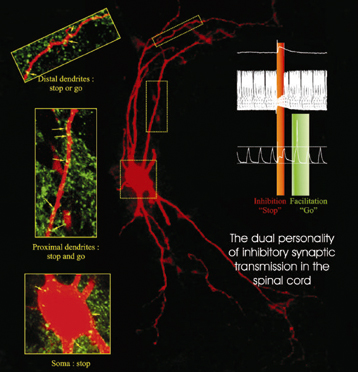
The above image comes from a CNRS press release about the “double personality” of inhibitor neurons. Oh, and yes, the press release is in French. Sorry ’bout that. CNRS maintains an English site as well, but it lags several weeks behind the French site (shocking, I know).

Basically, researchers have discovered a chemical basis for the function of inhibitor neurons—neurons that seem to play a role in disorders such as paralysis and epilepsy. According to the new findings, the firing of a neuron (“stop” and “go,” as it’s described in the figure above) depends on the concentration of chloride ions, which is in turn controlled by proteins in the surface of the neuron.

To my eye, the visualization (presumably of actual data) communicates its message quite clearly. First off, it capitalizes on existing color associations with “stop” (red) and “go” (green), but it also does a nice job of highlighting specific regions of the neuron (n.b. “stop or go” and “stop or go”). A lot of information in a small space. The little diagram on the upper left is far too small for me to make out, but I’m guessing that it contains information that I would find interesting were I able to read it.

The red-green color scheme also seems to correlate with the use of bioluminescent tags in various samples. The book Aglow in the Dark: The Revolutionary Science of Biofluorescence taught me a wee bit about this field. Fascinating stuff.

Stem Cell, Stem Cell, Burning Bright

Researchers at the University of Michigan Medical School and Japan’s University of Tsukuba have announced the ability to tag hematopoietic stem cells (HSCs) in living bone marrow—which means they managed to make the gene for green fluorescent protein (GFP) express itself only in HSCs and not in the surrounding cells. The glowing green blob in the image above represents a stem cell, all on its lonesome, in the midst of mouse bone (as opposed to mouse brains).

HSCs are the little folks responsible for forming blood cells and supporting the immune system, and they don’t survive too long outside of bone marrow, so it’s important to track their behavior where they’re most at home. The press release also links to a time-lapse QuickTime of the luminescent stem cell, although I should warn you that not much happens. The important thing to note is that the stem cell seems rather lonely; instead of hanging out with a bunch of pals, it (like the cheese) stands alone.

The technique of tagging cells with the gene for bioluminescence hasn’t been around too terribly long, but it provides a remarkably straightforward way of visualizing what’s happening at the cellular (or even sub-cellular) level. The image above gets the message across quite clearly.

Coincidentally, I recently picked up an impressive (and well-illustrated) tome that describes the development of bioluminescent tagging for use in research. Aglow in the Dark: The Revolutionary Science of Biofluorescence is written by two insiders in the field, who give an intimate introduction to the history of the topic, while providing truly enlightening (sorry) background to the scientific and technical challenges. Quite a fascinating little book.